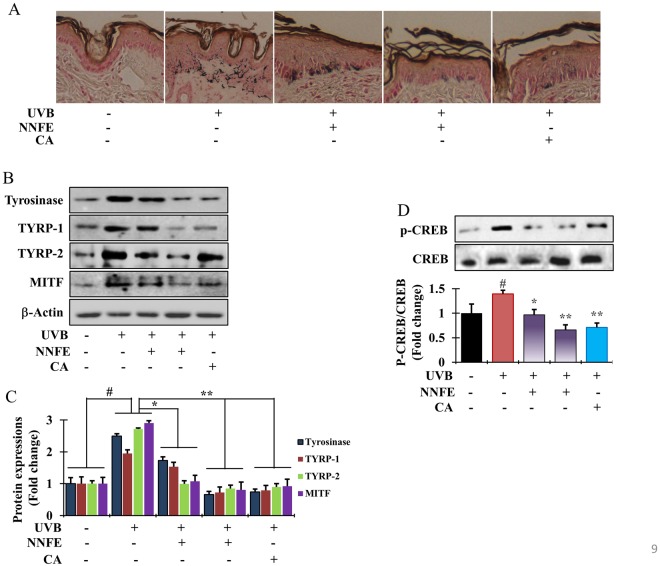
Figure 9.
Effects of NNFE on pigmentation of mouse skin exposed to UVB. (A) Fontana-Masson staining of dorsal skin sections from the mice shown in (Fig. 8B) reveals differences in melanin content. (B) After completing UVB treatment, dorsal skin was excised, homogenised, and assayed by western blot using antibodies against tyrosinase, TYRP-1, TYRP-2, and MITF. Equal protein loading was confirmed using anti-β-actin. Arb, arbutin. (C) Quantification and statistical analysis of the band intensities of tyrosinase, TYRP-1, TYRP-2, and MITF obtained by western blot analysis. *p < 0.05, **p < 0.01, versus the non-treated controls, Student’s t-test. (D) Phosphorylation of CREB confirmed by western blot analysis. NNFE, ethyl acetate fraction of N. nouchali flower extract; CA, caffeic acid.

