Figure 5.
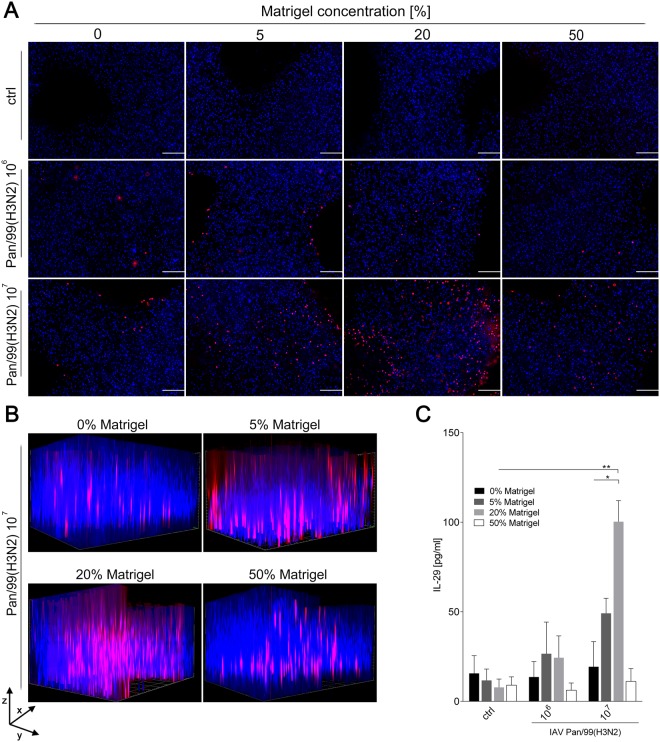
Distribution of Pan/99(H3N2) in cell-laden alginate/gelatin/Matrigel matrices and IL-29 release. 3D printed cell-laden alginate/gelatin constructs with varying Matrigel concentrations were infected with either 106 or 107 PFUs of Pan/99(H3N2) 24 hours after printing. (A,B) Fixed constructs were immunohistochemically labeled with anti-nucleoprotein antibody (red channel), and nuclear counter staining was performed using Hoechst stain (blue channel) 24 h after infection. Stained constructs were analysed by fluorescence microscopy by (A) top view (scale bar: 200 µm) or (B) Z-stack analysis to visualize spatial distribution of infected A549 cells (scanning depth 1000 µm, interval 15.12 µm, area 1800 × 1400 µm). (C) Supernatants of infected constructs were collected 24 h after Pan/99(H3N2) infection and release of IL-29 determined by ELISA. Results are shown as mean ± SEM of three independent experiments. *p < 0,05; **p < 0,01 compared to 0% Matrigel.

