Fig. 3.
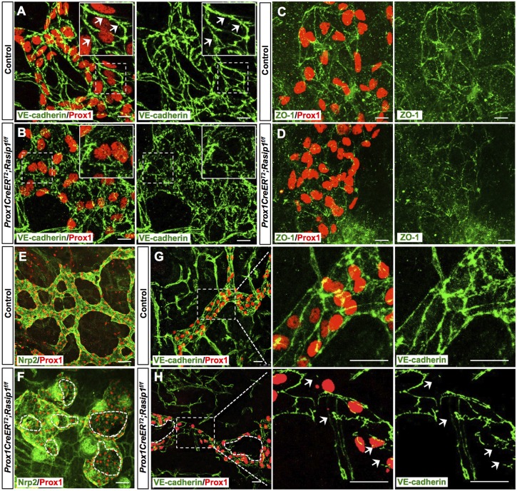
Disorganized cell junctions in dermal lymphatics of E14.5 Prox1CreERT2;Rasip1f/f embryos. (A,B) Whole-mount immunostaining of dermal lymphatic capillaries from E14.5 control (n=4) and Prox1CreERT2;Rasip1f/f (n=3) embryos. Insets are higher magnifications of selected regions (dashed boxes). Control lymphatics show zipper-like junctions as indicated by white arrows. Instead, obvious cell junctions are difficult to detect in Rasip1 conditional null littermates, as cell borders appear to be disorganized and diffuse. (C,D) Similar skin whole-mount immunostaining was performed in control (n=3) and Prox1CreERT2;Rasip1f/f (n=3) embryos but using antibodies against the tight junction ZO-1 and Prox1. In this case, junctions were also diffuse and defective, and levels of ZO-1 appeared to be reduced. (E-H) Whole-mount immunostaining of dermal lymphatic capillaries from E16 control (n=3) and Prox1CreERT2;Rasip1f/f (n=3) embryos. Dashed lines indicate open and broken lumens in F and H. Right panels show higher magnifications of the boxed regions in G and H. Arrows indicate the broken junctions in Rasip1 conditional null embryos. Scale bars: 10 μm in A-D; 50 µm in E,F; 25 µm in G,H.

