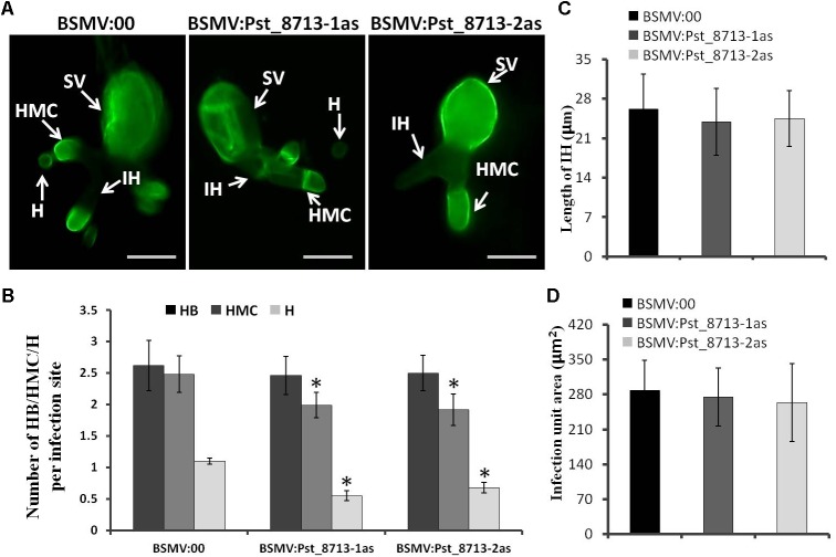
FIGURE 9.
Histological observation of fungal growth in wheat plants inoculated first with BSMV:00 or recombinant BSMV and then inoculated with isolate CYR32 (A) Fungal growth at 18 h after CYR32 inoculation. The fungal structure was stained with wheat germ agglutinin (WGA) and observed under a fluorescence microscope. Bars = 20 μm. (B) The average number of HB, HMC and H in HIGS plants infected by CYR32. (C) Hyphal length, which is the mean distance from the junction of the sub-stomatal vesicle of the hypha to the tip of the hypha, measured using DP-BSW software. (D) Infection area, the mean area of the expanding hyphae plus the host cells, was calculated using DP-BSW software (unit in ×103/μm2). All bars indicated means ± SE of three independent biological replicates with 50 unit areas per replicate, Asterisks indicate significant differences (P < 0.05) using Student’s t-test. SV, sub-stomatal vesicle; HMC, haustorial mother cell; IH, infection hypha, H, haustorium, and HB, hypha branch.

