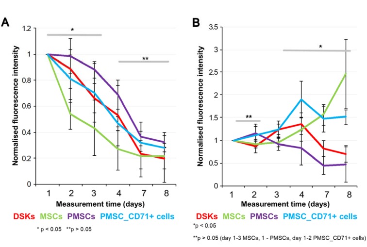
Figure 6. Cells fluorescence signal intensity in the full-thickness skin wound model. MitoTracker Deep Red labelled cells fluorescence intensities were estimated on 1st, 2nd, 3rd, 4th, 7th and 8th day after transplantation.
A. Intradermally injected dorsal skin keratinocytes (DSKs), mesenchymal stromal cells (MSCs), partially differentiated MSCs (PMSCs) and CD71 positive PMSC population (PMSC_CD71+ cells relative amount in the region of interest (ROI)
B. Intravenously injected DSKs, MSCs, PMSCs, PMSC_CD71+ cells in the ROI. Fluorescence intensity normalised to the first measurement. Error bars indicating standard deviation. n = 6 mice

