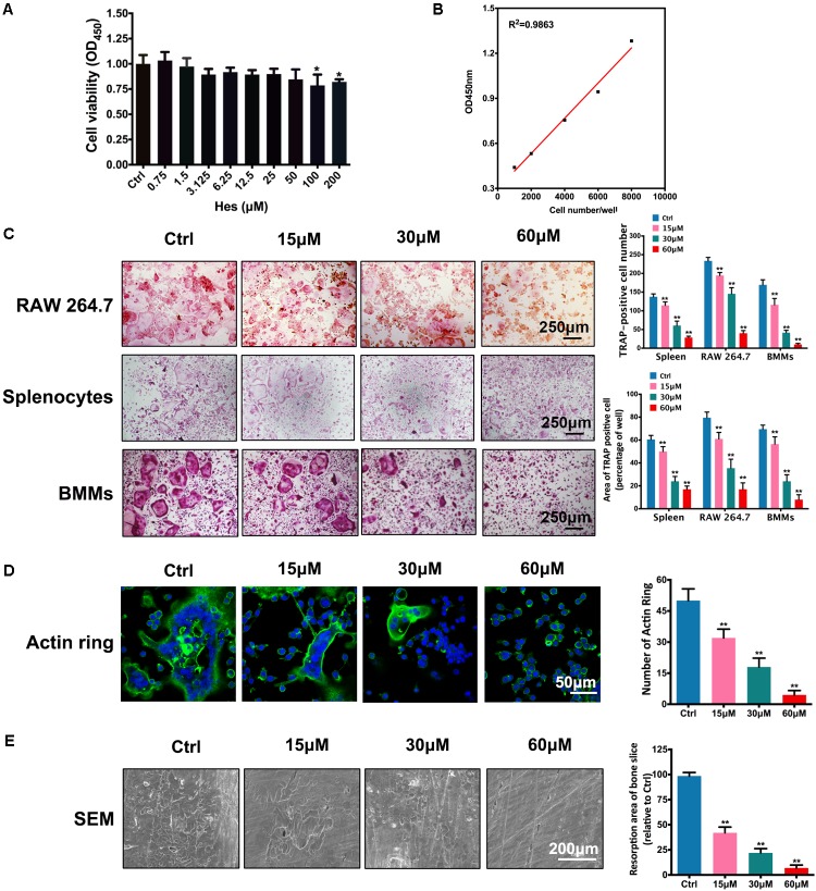
FIGURE 1.
Non-toxic Hes attenuated RANKL-induced osteoclast formation and function in vitro. (A) Cell viability of osteoclast precursors after Hes treatments for 24 h. (B) Linear correlation between OD values and cell numbers. (C) RANKL-induced osteoclastogenesis after Hes treatments in three types of preosteoclasts, RAW 264.7 cells, splenocytes, and BMMs. (D) Formation of RANKL-induced F-actin rings after Hes treatments. (E) Formation of RANKL-stimulated bone resorption pits after Hes treatments. ∗p < 0.05 compared with controls, ∗∗p < 0.01 compared with controls.

