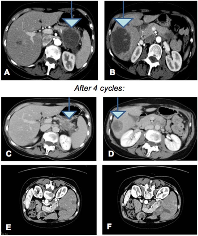
Figure 1.
Computed tomography scan of March 2014 showing peripancreatic lymph nodes and liver parenchyma with numerous secondary lesions (A and B). Computed tomography scan of July 2014 after 4 cycles of integrated therapy that showed a net reduction in pancreatic injury and secondary liver lesions (C and D). After 2 more cycles, removing oxaliplatin, the result was the disappearance of the nuanced liver (E and F).

