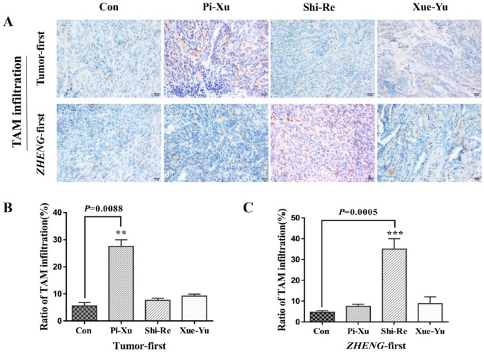
Figure 2.
Immunohistochemical analysis with a high-power (400×) microscope to assess TAM expression in subcutaneously transplanted pancreatic cancer tissues in “Tumor-first” and “ZHENG-first” models: (A) TAM expression was evaluated using a CD68 antibody; “Tumor-first” (top) and “ZHENG-first” (bottom). The CD68 positive staining ratio was quantitatively estimated to assess TAM infiltration; *P < .05. (B) “Tumor-first” model and (C) “ZHENG-first” model.

