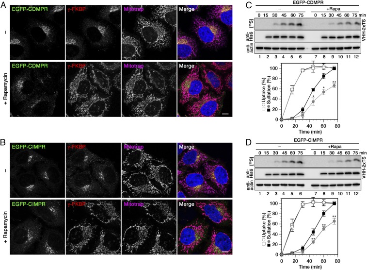
Fig. 6.
Retrograde transport of MPRs is reduced upon rapid depletion of AP-1. (A and B) HeLa-AP1ks cells stably expressing EGFP-CDMPR or -CIMPR after silencing endogenous γ-adaptin were treated with or without 500 nM rapamycin for 1 h and processed for fluorescence microscopy to detect EGFP-MPR, recombinant γ-FKBP, and Mitotrap. (Scale bars, 10 µm.) (C and D) HeLa-AP1ks cells stably expressing EGFP-CDMPR or -CIMPR after silencing endogenous γ-adaptin were labeled with [35S]sulfate for up to 75 min in the presence of 2 μg/mL VHH-2xTS with or without 500 nM rapamycin. Cell-associated nanobodies were isolated and subjected to immunoblot analysis (anti-His6) and autoradiography ([35S]). Aliquots of cell lysates were immunoblotted for actin. Quantitation of VHH-2xTS uptake and sulfation is shown in percent of the value without rapamycin after 75 min (mean and SD of three independent experiments; two-sided Student’s t test: *P < 0.05; **P < 0.01). Black squares, without rapamycin; gray circles, with rapamycin; open symbols, uptake; filled symbols, sulfation.

