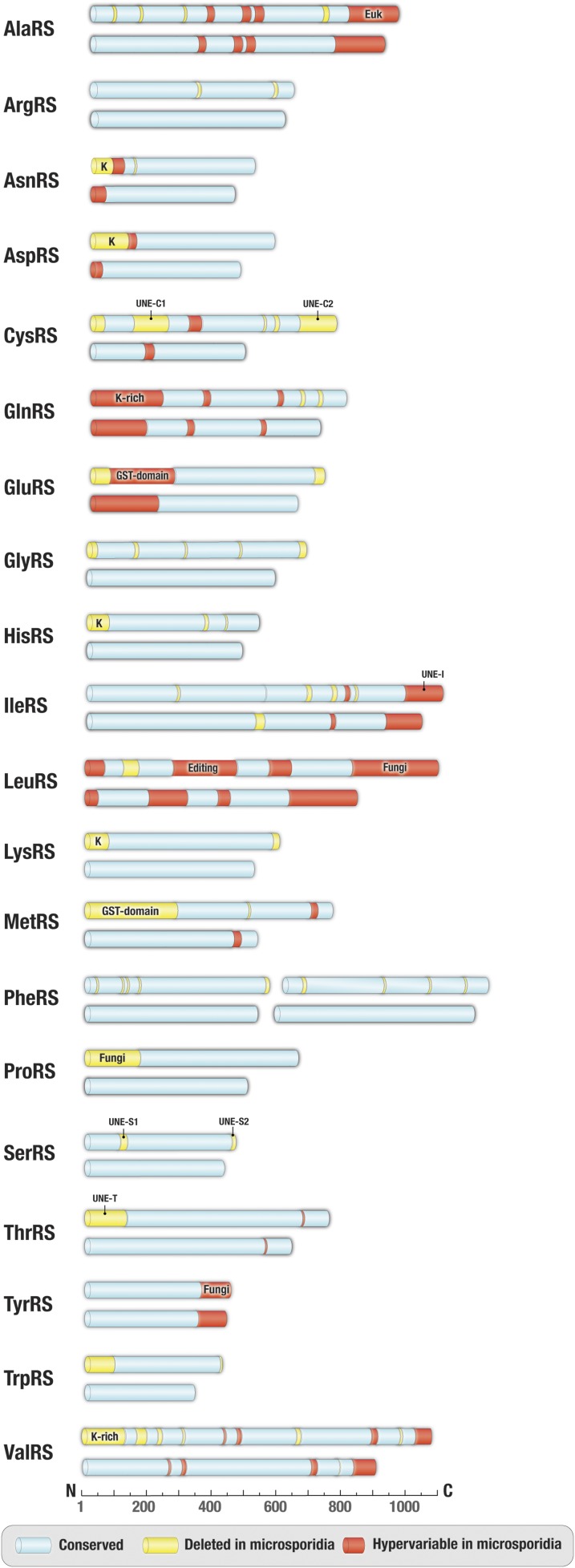
Fig. 1.
Apparent reductive evolution of microsporidian aminoacyl-tRNA synthetases. Schematic comparison of primary structures of aminoacyl-tRNA synthetases from microsporidian parasites (exemplified by V. culicis) and nonparasitic fungi (exemplified by S. cerevisiae). Protein segments highlighted in blue indicate conserved regions (sequence similarity >50%), protein segments present in yeast but deleted in microsporidia are shown in yellow, and segments with highly variable sequences in microsporidia (sequence similarity <35% between V. culicis and T. hominis) but are conserved in other eukaryotes are highlighted in red. Labels indicate appended domains, such as eukaryote-specific (Euk), fungi-specific (Fungi), GST-domain, UNE-domain, and lysine-rich (K-rich) domains.

