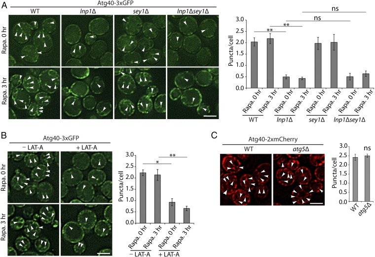
Fig. 5.
Fewer Atg40 puncta are observed in the cell interior in the lnp1Δ mutant and in LAT-A–treated cells. (A) Cells expressing Atg40-3xGFP were untreated (0 h) or treated for 3 h with rapamycin. Arrowheads mark Atg40 puncta located in the cell interior. (A, Right) Quantitation of Atg40-3xGFP puncta in the cell interior. Error bars represent SEM from three separate experiments. Puncta were quantitated from the following number of cells: WT at 0 h, n = 379; WT at 3 h, n = 454; lnp1∆ at 0 h, n = 511; lnp1∆ at 3 h, n = 325; sey1∆ at 0 h, n = 361; sey1∆ at 3 h, n = 288; lnp1∆ sey1∆ at 0 h, n = 306; lnp1∆ sey1∆ at 3 h, n = 413. **P < 0.01, Student’s t test; ns, nonsignificant. (B) Cells were treated with rapamycin with or without 100 µM LAT-A as described in Materials and Methods. Arrowheads mark Atg40 puncta located in the cell interior. (B, Right) Quantitation of Atg40-3xGFP puncta in the cell interior. Error bars represent SEM from three separate experiments. Puncta were quantitated from the following number of cells: −LAT-A at 0 h with rapamycin, n = 348; −LAT-A at 3 h with rapamycin, n = 292; +LAT-A at 0 h with rapamycin, n = 420; +LAT-A at 3 h with rapamycin, n = 314. *P < 0.05, **P < 0.01, Student’s t test. (C) WT and atg5Δ mutant cells were treated for 3 h with rapamycin. Arrowheads mark the Atg40 puncta located in the cell interior. (C, Right) Quantitation of Atg40-2xmCherry puncta in the cell interior. Error bars represent SEM from three separate experiments. Puncta were quantitated from the following number of cells: WT, n = 319; atg5∆, n = 280; ns, nonsignificant. (Scale bars, 3 μm.)

