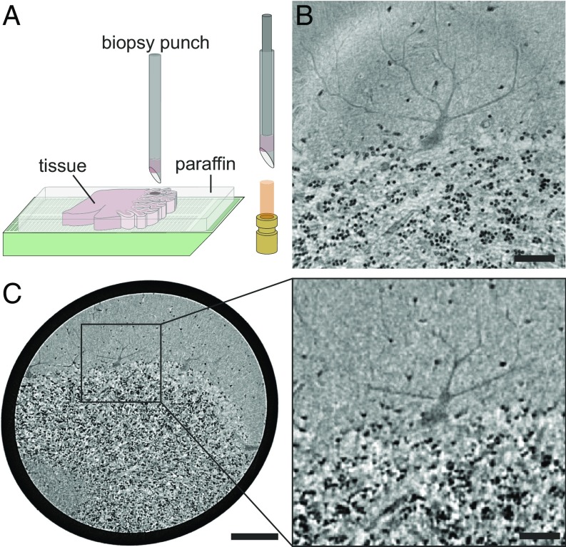
Fig. 1.
Virtual histology of human cerebellum. (A) Sample preparation for tomographic experiments. Biopsy punches were taken from paraffin-embedded human cerebellum and placed into a Kapton tube for mounting in the experimental setups. (B) Transverse slice through the reconstructed volume of the synchrotron dataset revealing the interface between the low-cell molecular and the cell-rich granular layer, including a cell of the monocellular Purkinje cell layer. (C, Left) Corresponding slice of the laboratory dataset (same plane, same specimen as in B), showing the larger volume accessible by the laboratory setup while maintaining the resolution required for single-cell identification. (C, Right) Magnified view of the region marked by the rectangle in C, Left corresponding to the field of view of the synchrotron dataset in B. [Scale bars: 50 m (B and C, Right) and 200 m (C, Left).]

