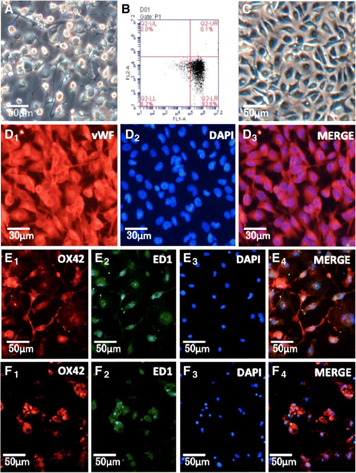
Fig. 2.
Characterization of rat retinal microglia and RMECS. A: Cultures of primary isolated microglia. B: The purity of isolated microglia detected by low cytometry through its specific surface marker CD11b is 93.6%. C: Culture of RMECs. D1-D3: Immunofluorescent detection of RMECs marked by vWF, DAPI and both. E1–E4: Normal microglia are static. F1 − F4: Exposure to 100 ng/mL LPS for 24 h altered the morphology of the microglia from a ramified state with long processes to the activated state with an amoeboid appearance. E1-E4, F1-F4 has been published in our previous research. Abbreviations: vWF, von-Willebrand factor; LPS, lipopolysaccharide. (Page9–10)

