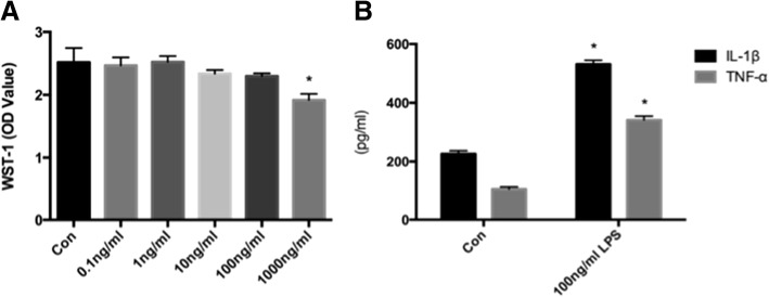
Fig. 3.
a Concentration-dependent effects of LPS on the viability of microglia. Microglia were exposed to LPS (0, 0.1, 1, 10, 100, or 1000 ng/mL) for 24 h. Microglial viability was measured with WST-1 reagent. LPS at 1000 ng/mL significantly reduced the viability of microglia. This part has been published in our previous research. b Effects of LPS on TNF and IL-1beta secretion. Microglia were exposed to LPS (0 and 100 ng/mL) for 24 h. The TNFα and IL-1beta concentration in the supernatant was measured using an ELISA kit. LPS at concentrations of 100 ng/mL significantly increased the production of TNF and IL-1beta. Results are means ± SD (n = 3 per group). *P < 0.05 vs Con using one-way ANOVA. Abbreviations: Con, microglia without LPS; WST-1, water soluble tetrazolium-1; LPS, lipopolysaccharide; TNFα, tumor necrosis factor; IL-1β, interleukin 1β. (Page 10, paragraph3–4)

