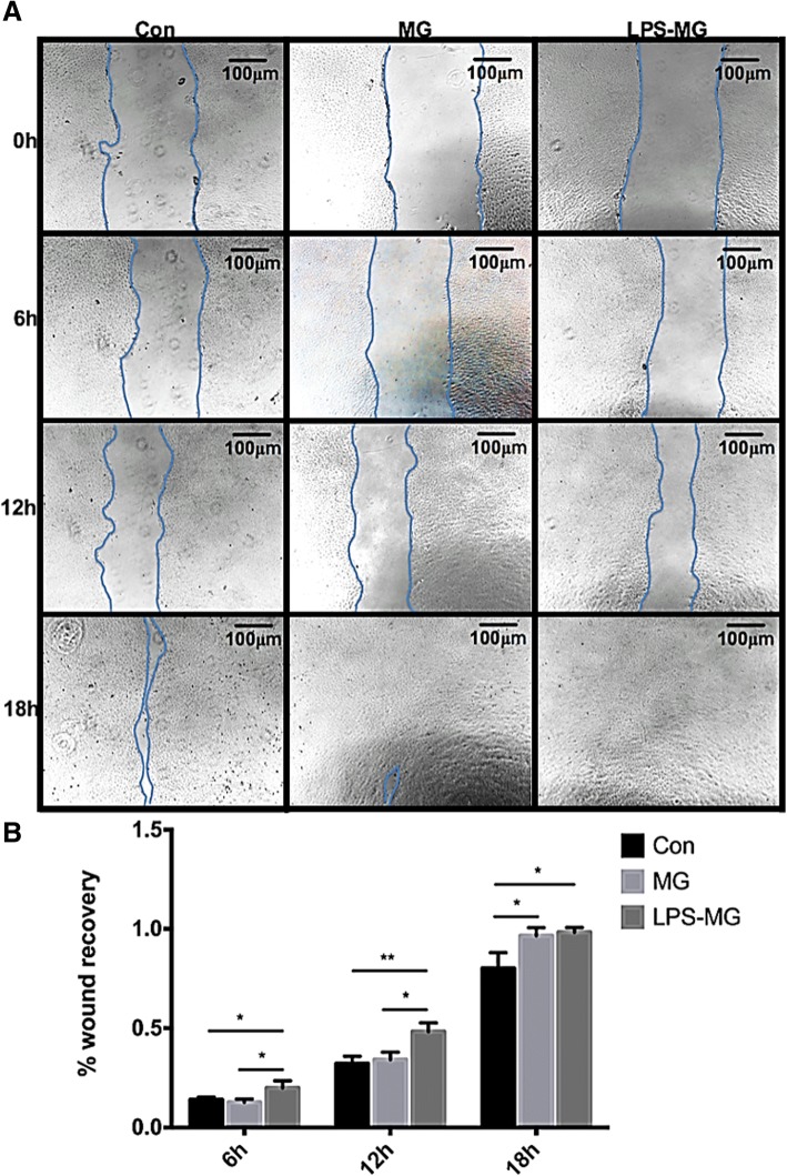
Fig. 6.
Effects of static or activated microglia on migration of RMECs. a One representative experiment of three different groups at 0 h, 6 h, 12 h, 18 h. b Wound recovery percentage analyzed by ImageJ. We see static microglia significantly stimulated wound recovery at 18 h, at the same time, activated microglia significantly stimulated wound recovery at 6 h, 12 h and 18 h. Each bar graph indicates means ± SD of three independent experiments. *Significant difference in results between the two compared groups. *P < 0.05 using one-way ANOVA. **P < 0.01 using one-way ANOVA. Abbreviations: Con, control RMECs (i.e., cultured without microglia); MG, REMCs co-cultured with static microglia; LPS-MG, REMCs co-cultured with activated microglia; LPS, lipopolysaccharide. (Page 11, paragraph 4)

