Fig. 1.
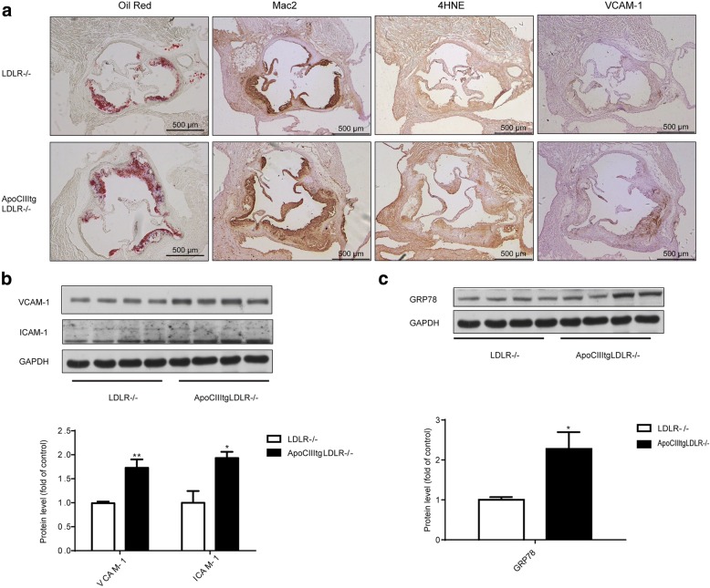
Increased inflammation, ER stress and oxidative stress in aorta of ApoCIIItgLDLR−/− mice comparing to LDLR−/− mice. (a) Representative images of Oil Red O (ORO) stained aortic roots and immunohistochemical staining of aortic sinus sections of Mac2, 4HNE and VCAM-1 expression in LDLR−/− and ApoCIIItgLDLR−/− mice. (b) Representative Western blot images of VCAM-1 and ICAM-1 protein expression in aortas of LDLR−/− and ApoCIIItgLDLR−/− mice and the protein quantification by densitometry (n = 4). (c) Representative Western blot images of GRP78 protein expression in aortas of LDLR−/− and ApoCIIItgLDLR−/− mice and the protein quantification by densitometry (n = 4). Values are expressed as mean ± SEM, *p < 0.05 **p < 0.01

