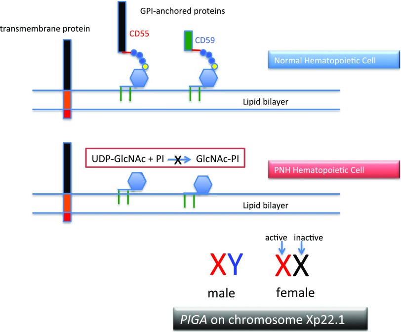
Figure 1.
The molecular basis of PNH. Normal hematopoietic stem cells express both transmembrane and GPI-anchored proteins (top). PNH stem cells express transmembrane proteins normally but fail to express GPI-APs because the first step in synthesis of the anchor is inactivated because the gene (PIGA) that encodes the enzyme that is required for transfer of the nucleotide sugar (UDP-GlcNAc) to phosphatidylinositol is mutant (middle). Of the more than 25 genes involved in synthesis of the GPI anchor, only PIGA is located on the X-chromosome (all others are autosomal). Location on the X-chromosome accounts for the observation that essentially all cases of PNH are due to somatic mutation of PIGA because inactivation of only 1 allele is required to produce the PNH phenotype as males have 1 X-chromosome and in females only 1 of the 2 X-chromosomes is active in somatic tissues (bottom). UDP, uridine diphosphate.

