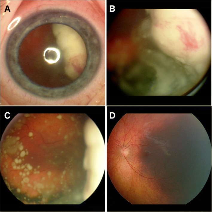
Fig. 1.
External (a) and fundus images (b,c) OD demonstrating solid white, predominantly endophytic retinal tumor with preretinal neovascularization on its surface (b) and extensive vitreous seeds (b, c). Fundus image OS (d) demonstrating broad-based flat melanotic area of choroid extending from the subfoveal region temporally

