Figure 5.
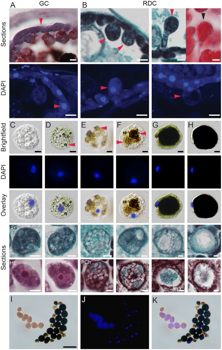
Development of digestive cells in spider mite midgut. (A) Brightfield microscopy (cryosection, Oil Red O staining) and fluorescence microscopy (paraffin sections, DAPI staining) of generative cells (GC) forming midgut epithelium. (B) Brightfield microscopy (paraffin sections, Fast Green and Safranin O, and Hematoxylin and Eosin staining) and fluorescence microscopy (paraffin sections, DAPI staining) of residual digestive cells (RDC) as they grow into the midgut lumen and detach from the midgut epithelial layer. (C–H) Digestive cells (DC) at various stages of development. DCs were dissected from the midgut lumen of female spider mites, stained using DAPI, and observed using brightfield and fluorescence microscopy. Lower panels, paraffin sections of DCs from different staining preparation: FG, Fast Green/Safranin O; HE, Hematoxylin/Eosin and Pch, Pentachrome. (C) Stage 1, the early stage of DC development with clear but numerous vesicles and large nucleus. (D) Stage 2, DC starting to uptake midgut lumen and plant pigments. Arrowhead marks colored vesicles within DC. (E,F) Stage 3, colored vesicles enlarge. (G) Stage 4, a large vesicle with dark content is surrounded by a thin layer of cytoplasm. Numerous small vesicles are still visible in cytoplasm, nucleus is displaced to the periphery. (H) Stage 5, the terminal stage of DC development characterized by a dense dark brown/black vesicle, a very thin crescent of cytoplasm and condensed nucleus on side of a DC. (I–K) Brightfield and fluorescence microscopy of spider mite feces. Feces are represented by guanine pellets and DCs at various stages of development. There is no apparent sorting of DCs based on developmental stage and amount of waste products accumulated. Guanine pellets readily absorb DAPI and also exhibit autofluorescence. Scale bars: (A): 10 μm; (B–H): 5 μm; (I–K): 50 μm.

