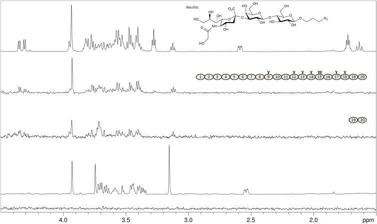
Fig. 3.
FH binds the nonhuman Neu5Gc variant of sialic acid. From top: Proton 1D reference spectrum of Neu5Gcα2-3Galβ1-4Glcβ-propyl-N3 (glycolyl-3’SL-ProN3, chemical structure shown; Neu5Gc differs only by one additional OH-group in the acetyl side chain from the Neu5Ac Sia-variant in 3’SL); STD NMR difference spectrum of 2 mM glycolyl-3’SL-ProN3 with 8 μM of FH; STD NMR difference spectrum of 2 mM glycolyl-3’SL-ProN3 with 50 μM of FH19–20; Proton 1D reference spectrum of Neu5GcαOMe; STD NMR difference spectrum of 2 mM Neu5GcαOMe with 8 μM of FH. The structures of FH and FH19–20 are shown schematically with glycosylation sites highlighted as “Y”. Magnetization transfer from the Neu5Gc-trisaccharide is observed for all three pyranose rings but not for the propyl linker protons (resonances at 1.7 ppm and 3.25 ppm), while no transfer is observed for the monosaccharide (bottom spectrum).

