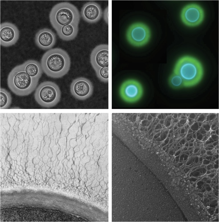
Fig. 1.
Cryptococcus neoformans capsule and cell wall. Clockwise from upper left: Negative stain of cryptococcal cells with India ink; immunofluorescence montage of capsule-induced cells stained with Calcofluor white (blue, stains cell wall) and antibody to the major capsule polysaccharide (green); quick freeze deep-etch electron micrograph (EM) of a portion of the plasma membrane (lower left region of the micrograph), bounded by the two layers of the cell wall and the associated capsule fibers (extending up and to the right); transmission EM of cryptococcal cell edge with capsule fibers extending upwards from the cell wall.

