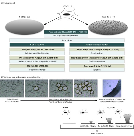
Figure. Summary of Materials and Methods Used in the Study.
A, Study protocol. B, Technique used for laser capture microdissection (LCM). ECM indicates extracellular matrix; EnMT, endothelial to mesenchymal transition; FECD-DM, Fuchs endothelial corneal dystrophy membrane; mRNA, messenger RNA; N-DM, normal Descemet membrane; PI, propidium iodide; RT-PCR, reverse-transcription polymerase chain reaction.

