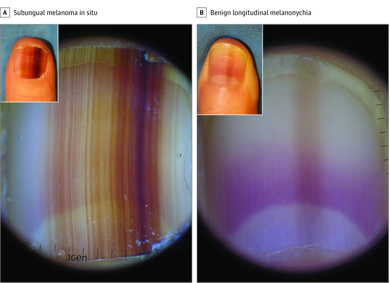
Figure 2. Dermoscopic and Clinical Manifestations of Subungual Melanoma In Situ and Benign Longitudinal Melanonychia.
A, Subungual melanoma in situ showing a 10-mm band with asymmetry, border fading, and multicolor pigmentation (dark and light brown). Inset, Clinical appearance of the involved nail. B, Benign longitudinal melanonychia showing a light brown band with a width of less than 2 mm on dermoscopic examination. Inset, Clinical appearance of the involved nail. Scale units are in millimeters.

