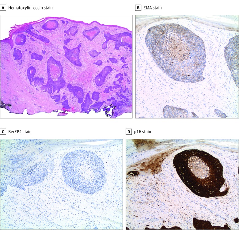
Figure 2. Histopathologic and Immunohistochemical Analyses of Basaloid Squamous Cell Carcinoma Tumors at Baseline.
A, Infiltrating islands of basaloid cells in a fibrotic stroma extending to the deep dermis (hematoxylin-eosin, original magnification ×20). B, Positive epithelial membrane antigen (EMA) stain findings. C, Negative BerEP4 stain findings. D, Positive p16 stain findings (original magnification ×200).

