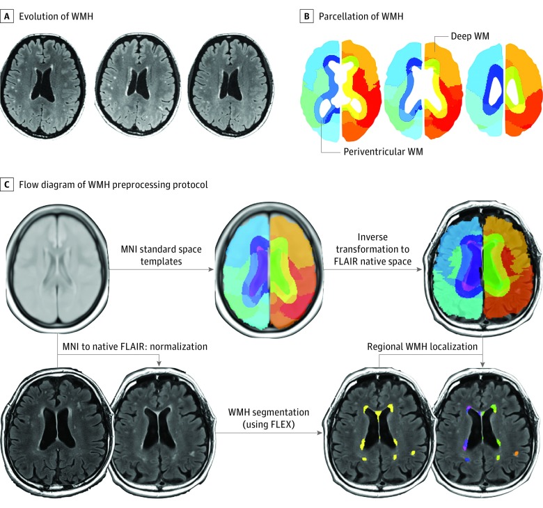
Figure 1. Segmentation of White Matter Hyperintensity Lesions (WMH) in Patients With Reversible Cerebral Vasoconstriction Syndrome.
A, Typical evolution of WMH in a patient with reversible cerebral vasoconstriction syndrome. Left panel, day 16; middle panel, day 29; right panel, day 84. B, Parcellation of WMH into periventricular (≤13 mm from ventricles) and deep (>13 mm from ventricles) lesions. C, The flow diagram of the WMH preprocessing protocol used in this study. The isotropic 3-dimension fluid-attenuated inversion recovery (FLAIR) images were used to perform automatic WMH segmentation with the FLAIR lesion segmentation toolbox (FLEX). An inverse transformation matrix derived from the nonlinear registration between native T2 images and FLAIR templates in MNI standard space was used to transform the brain atlas. Individual brain atlases were used for WMH localization.

