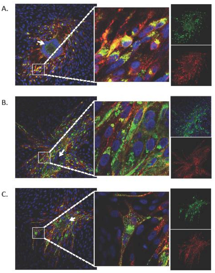
Fig. 5. Localization of VZV gM in C-terminal mutants.
The cellular localization of VZV gM in rVOka (A), the gM-double YxxΦ (B) and gM triple mutant (C) viruses was evaluated by confocal immunofluorescence of VZV-infected fibroblasts at 48 hours post infection using an anti-gE (red signal) and rabbit anti-gM (green signal). Cell nuclei are stained using Hoechst (blue). Inset panel (middle) shows high mag of small white square in left panel. Single color images for red and green signal are shown in the far right side. Arrows are described in Results.

