. 2018 Aug 15;7:e38325. doi: 10.7554/eLife.38325

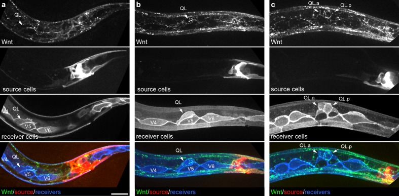
Figure 2. Endogenously tagged Wnt/EGL-20 localizes to responding QL neuroblasts that do not directly contact Wnt source cells.

(a–c) Maximum intensity projections showing Wnt/EGL-20::mNG protein (green), plasma membranes of EGL-20 source cells marked by Pegl-20>2x mKate2::PH (red), and responding Q neuroblast and seam cell membranes marked by Pwrt-2>2x mTurq2 (blue) in L1 larvae during QL polarization (a), early QL migration over the seam cell V5 (b) and the onset of QL descendant migration after the first cell division (c). Images are oriented with anterior to left and dorsal to top, scale bar = 10 μm.

Figure 2.

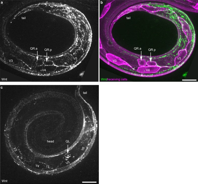
Figure 2—figure supplement 1. Wnt/EGL-20::mNG localization in QR neuroblast descendants and seam cells.

Figure 2—figure supplement 1.

(a–b) Maximum intensity projections of sub-surface optical sections on the right side of an L1 stage larva showing bright Wnt/EGL-20::mNG punctae (green) that localize to anteriorly migrating QR neuroblast descendants and the posterior ends of seam cells V3-V6 (magenta). (c) Maximum intensity projection of surface optical sections on the left side of and L1 larva highlighting bright Wnt/EGL-20::mNG punctae that localize to the QL neuroblast and seam cells V3-V6. QL has migrated over V5 but has not yet divided. Animals are curled with the tail to upper right, scale bars = 10 μm.