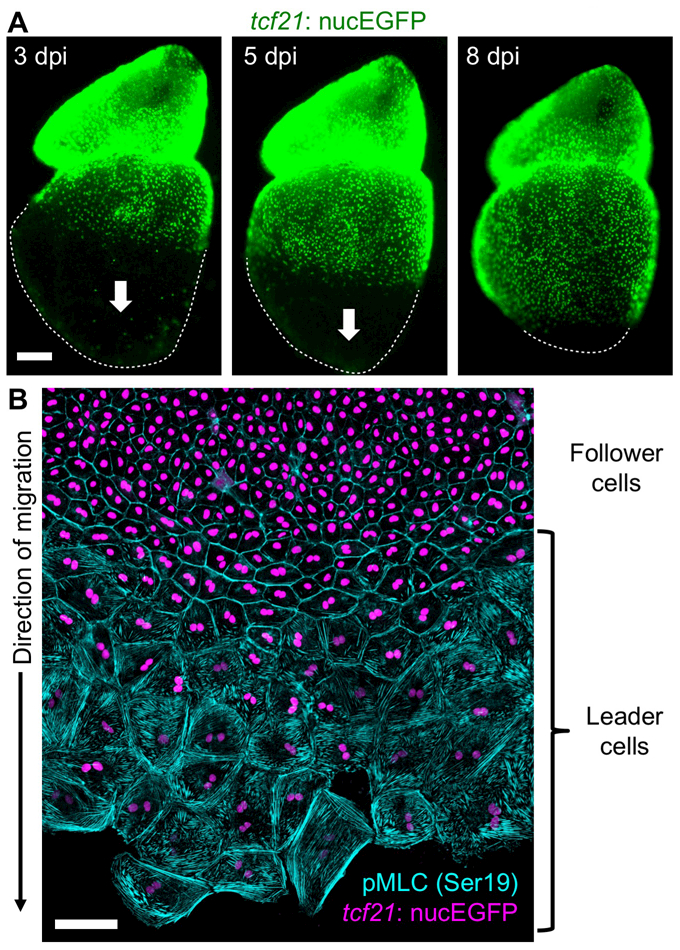
Figure 3. Ex vivo epicardial regeneration.

(A) Whole-mount images of explanted zebrafish heart showing epicardial regeneration along the ventricular surface in a base-to-apex direction (arrows). tcf21:nucEGFP visualizes epicardial cell nuclei (green). dpi, days post Mtz incubation. (B) The epicardium generates a leading edge of large, multinucleate (leader, bottom) cells during migration ex vivo. Trailing (follower, top) cells are small and mononucleate. Epicardial nuclei are indicated in violet (tcf21:nucEGFP) and phosphorylated myosin light chain 2 (Ser19), an indicator of mechanical tension, is cyan.
