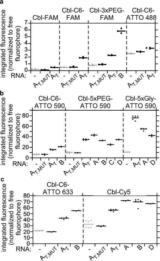
Figure 2.

Cbl riboswitch RNAs induce fluorescence turn-on in Cbl-fluorophore probes in vitro. The fluorescence intensity of Cbl-fluorophore probes in the presence or absence of different RNAs was quantified as in Figure 1d and normalized relative to the intensity of the free fluorophore. RNAs A, B, C and D are variants of Cbl-binding riboswitch sequences, where AT refers to a truncated version of A with linker region J1/3 and stem-loop P/L13 deleted (see also Figure 1b). AT,MUT contains four point mutations in the Cbl-binding site (see Figure 1b for the position of these residues). (a) Probes with fluorescence in the green wavelength range (n=3 independent measurements); (b) probes with fluorescence in the red wavelength range (from left to right, n=9, 3, 6, 3, 6, 3, 6, 3, 3, 3, 3, 12, 6, 6, 3 independent measurements), (c) probes with fluorescence in the far red wavelength range (from left to right, n=6, 3, 6, 3, 6, 3, 6, 3, 6, 3). See Supplementary Table 4 for a summary.
