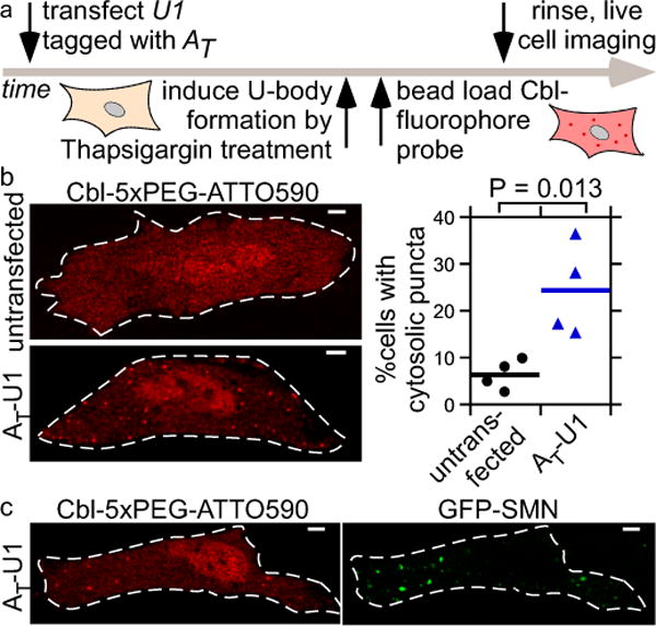
Figure 5.

Monitoring cytosolic U-bodies via AT-tagged U1. (a) After transient transfection of AT-U1, U-bodies were induced by thapsigargin treatment followed by live cell microscopy. (b) Cbl-5xPEG-ATTO 590 localization to cytosolic puncta in thapsigargin-treated HeLa cells is more likely when AT-U1 was transfected (AT-U1: mean from 4 independent experiments/326 cells; untransfected: mean from 4 independent experiments/677 cells). One way ANOVA (95% confidence limit), post hoc test (Tukey HSD). (c) Cytosolic puncta in thapsigargin-treated cells producing AT-U1 RNA co-localize to GFP-SMN U-bodies. 3 independent experiments, 10 cells. Scale bar = 5 μm.
