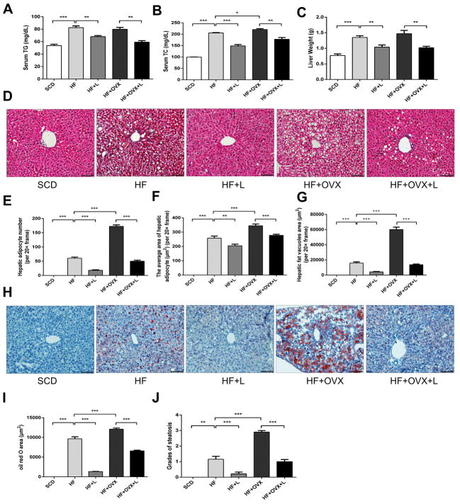
Figure 3. Effects of knee loading on hepatic steatosis.
A) Serum triglyceride levels. B) Serum cholesterol levels. C) Liver weight. D) Representative HE-stained sections of liver tissues (magnification 200×; and scale bar=100μm). E-G) Measurement of the hepatic adipocyte number, average area of a hepatic adipocyte, and area of hepatic fat vacuoles. H) Oil Red O stained sections of liver tissue (magnification 200×; and scale bar=100μm). I) Measurement of Oil Red O stained area. J) Grades of steatosis. All values were expressed as mean±SEM (n=10 mice per group). The asterisks (*, ** and ***) represent P<0.05, P<0.01, and P<0.001, respectively.

