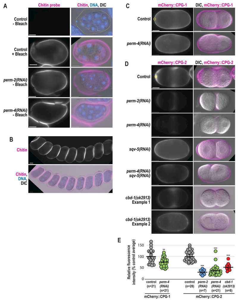
Figure 5. PERM-2 and PERM-4 promote structural integrity of the vitelline layer.

(A,B) Immunofluorescence of fixed embryos stained with a rhodamine-conjugated chitin-binding probe to mark the eggshell (magenta) and DAPI to mark DNA (blue). (C,D) Widefield fluorescence and DIC images of live embryos from control, RNAi-treated, and cbd-1(ok2913) mutant worms that express mCherry::CPG-1 (C) or mCherry::CPG-2 (D). n=number of embryos. Scale bars, 10um. (E) Quantification of relative fluorescence intensity of eggshell markers within a region of interest (ROI, yellow circles) normalized to the control average. Horizontal black bars represent the mean, vertical black bars the standard error of the mean (SEM). **, p=0.0033;***, p<0.0001 compared to controls by unpaired student’s t-test with unequal variance.
