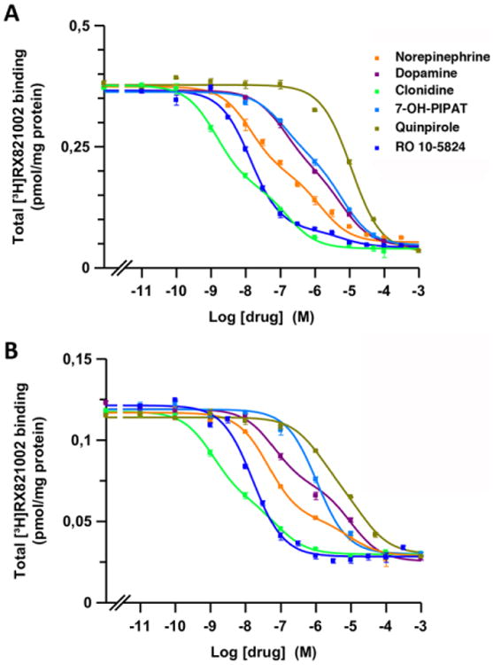
Figure 2. Radioligand binding of dopaminergic and adrenergic ligands to α2 adrenoceptors in brain tissue.

Representative competition curves of α2 adrenoceptor antagonist [3H]RX821002 vs. increasing concentrations of free competitors (NE, DA, clonidine, quinpirole, 7-OH-PIPAT and RO-105824) in sheep brain cortical (A) and striatal (B) membranes. Experimental data were fitted to the two-state dimer receptor model equations, as described in the Materials and Methods section. Values are mean ± S.E.M. from a representative experiment (n = 3) performed in triplicate.
