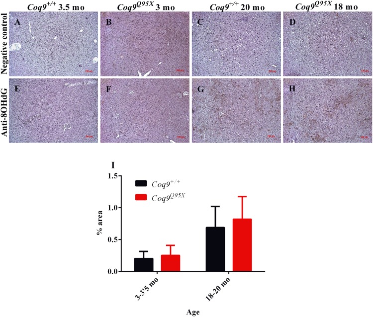
Figure 5.
Inmunohistochemistry against 8-OHdG as a marker of oxidative damage. (A–D) Representative negative control of the liver at 3–3.5 month of age in Coq9+/+ (A) and Coq9Q95X mice (B); and at 18–20 month of age in Coq9+/+ (C) and Coq9Q95X (D) mice. (E–H) Representative anti-8OHdG immunohistochemistry of the liver at 3-3.5 month of age in Coq9+/+ (E) and Coq9Q95X mice (F); and at 18–20 month of age in Coq9+/+ (G) and Coq9Q95X (H) mice. Data information: Scale bars: 100 µm. (I) Percentage of 8OHdG positive signal in the images. Results correspond to the mean ± SD, as determined by the software ImageJ. Coq9+/+ mice n = 3; Coq9Q95X mice n = 3, at each age.

