Figure 4.
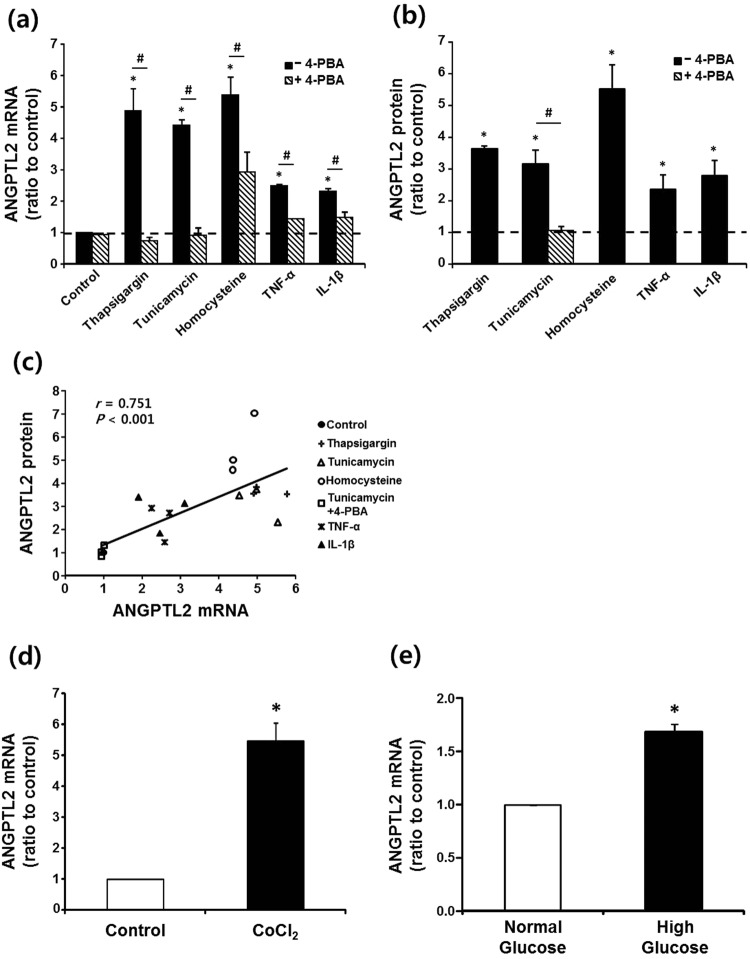
Induction of ANGPTL2 in human adipocytes. (a,b) Up-regulation of ANGPTL2 by ER stressors and proinflammatory cytokines. Fully differentiated human adipocytes (n = 3) were incubated in serum-free media for 24 hours with or without (control) various stressors such as thapsigargin (500 nM), tunicamycin (2 μg/mL), homocysteine (4 mM), TNF-α (10 ng/mL), and IL-1β (10 ng/mL). Effect of chemical chaperon was examined by adding 4-PBA (500 μM) to the culture media 2 hours prior to the treatment with stressors. ANGPTL2 mRNA level was measured by qPCR using 36B4 as the reference gene (a) and secreted ANGPTL2 protein was assessed by measuring its concentration in the culture media by ELISA (b). *p < 0.05 vs. control by ANOVA with Tukey test, #p < 0.05 vs. without 4-PBA by paired t-test. Dotted line represents ANGPTL2 mRNA or protein level at control state without stimulation. (c) Correlation between ANGPTL2 mRNA expression and ANGPTL2 protein secretion in differentiated human adipocytes. The correlation coefficient (r) between the measurements in experiments described above in A & B was calculated using Spearman’s correlation. (d,e) Effects of hypoxia or glucose on ANGPTL2 mRNA expression. (d) Differentiated human adipocytes were incubated in serum-free media for 24 hours with or without (control) a chemical hypoxic inducer CoCl2 (100 μM). *p < 0.05 vs. control by paired t-test. (e) Cells were incubated in the media containing either 5.5 mM (normal glucose) or 25 mM glucose (high glucose). *p < 0.05 vs. the normal glucose condition by paired t-test.

