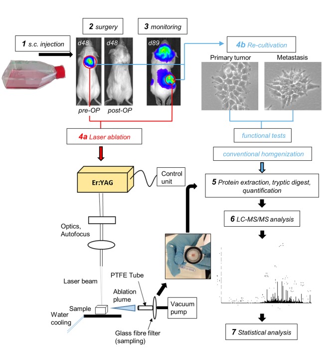
Figure 1.
Experimental setup. Human neuroblastoma LAN-1-Luc2/mCherry cells were subcutaneously injected into immunodeficient rag2−/− mice and surgically resected at a xenograft tumor size of ~1 cm3. Before and after surgery, mice were analyzed by bioluminescence imaging (BLI) to demonstrate the absence of detectable metastases at the time of surgery. Primary tumor cells were retrieved for in vitro expansion and establishment of the subline LAN-1-PT. Regular post-operative BLI scans were used to monitor the outgrowth of distant metastases. These lesions were either subjected to IR-laser ablation of proteins and subsequent proteome analysis or recovered for in vitro-cultivation. The resulting metastatic sublines LAN-1-Met-L and LAN-1-Met-O were functionally characterized and prepared for proteome analysis.

