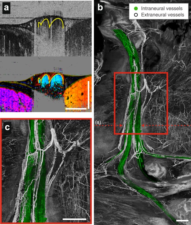
Figure 4.
Wide-field three-dimensional angiography with fascicular segmentation in a native nerve. (a) Cross-sectional angiography and vectorial birefringence imaging together highlight vessels and identify those within the nerve fascicle. (b) Fascicular vessels are colorized green in en face angiographic projections that reveal the vascular networks in the healthy peripheral nerve and surrounding tissues. (c), Expanded view of the region marked by the red rectangle in (b). Scale bars = 1 mm.

