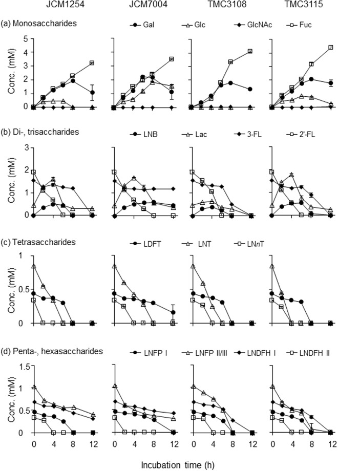
Figure 3.
In vitro HMO degradation behaviour of four B. bifidum strains. Each strain was cultured in HMOs-containing basal media in triplicate, and samples were collected at the indicated time points (see Fig. S2). The sugars in the culture supernatants were labelled with 2-AA and analysed by HPLC, as described in the Methods section. Note that LNB was not accurately quantified due to its heat lability. Concentrations of (a) monosaccharides (Fuc, Gal, Glc, and GlcNAc), (b) di- and trisaccharides (2′-FL, 3-FL, Lac, and LNB), (c) tetrasaccharides (LNT, LNnT, and LDFT), and (d) penta- and hexasaccharides (LNFP I, LNFP II/III, LNDFH I, and LNDFH II) are shown. The data are means ± SD of the labelled sugars obtained from three separate cultures.

