Fig. 1.
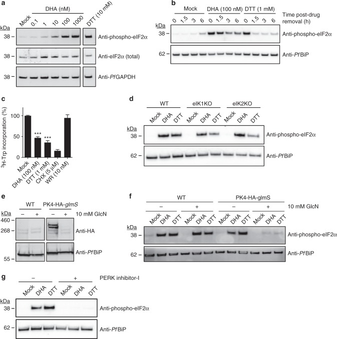
DHA activates an unfolded protein response, mediated by PK4. a, b Trophozoites (a 3D7, b Cam3.II_rev) were treated with 0.1% DMSO (mock), DHA or DTT for 90 min and harvested immediately (a) or washed and returned to culture for the indicated amount of time (b) before lysates were subjected to Western blot analysis and membranes probed for phosphorylated-eIF2α. c Trophozoites (Cam3.II_rev) were incubated with indicated compounds for 1 h and protein synthesis measured. Error bars represent s.e.m. ***P < 0.001 between mock and DHA or DTT treated samples (n = 5 (Mock); n = 4 (DTT); n = 3 (DHA, WR99210, CHX)), ANOVA. d eIK1 and eIK2 knock-out trophozoite-stage parasites (in a 3D7 background) were treated with 0.1% DMSO (mock), 1 μM DHA or 1 mM DTT for 60 min and lysates were subjected to Western blot analysis and probed for phosphorylated-eIF2α. e, f PK4-HA-glmS (in a 3D7 background) or WT (3D7) trophozoite-infected RBCs were treated with 10 mM glucosamine (GlcN) from the ring stage of the previous cycle and lysates were subjected to Western blot analysis, probing with anti-HA (e), or parasites were treated with 0.1% DMSO (mock), 100 nM DHA or 1 mM DTT for 60 min and lysates were subjected to Western blot analysis, probing for phosphorylated eIF2α (f). g Trophozoites (Cam3.II_rev) were treated with 20 μM PERK Inhibitor-I for 3 h, followed by a further hour in 0.1% DMSO (mock), 1 μM DHA or 1 mM DTT and lysates were analysed by Western blot for phosphorylated-eIF2α. Loading controls, PfGAPDH or PfBiP. All blots are representative of at least three independent experiments. Additional details in Supplementary Fig. 1

