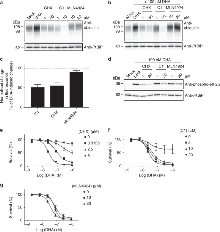
Fig. 3.
Compounds that inhibit protein polyubiquitination and protein damage antagonise the activity of DHA. a, b Trophozoites (3D7) were subjected to the indicated treatment for 6 h before analysis of ubiquitinated proteins. Samples treated with MLN4924 were incubated for an additional 3 h prior to DHA treatment. Blots are representative of 3–4 independent experiments (see Supplementary Fig. 3 for biological replicates). c Normalised maximum change in GFP fluorescence, measured by flow cytometry after GFP-DD trophozoites were incubated with 20 μM of the indicated compounds for 3 h, prior to exposure to DHA for 3 h. Dotted line represents 100%, the maximum decrease in fluorescence with DHA treatment. Error bars represent s.e.m. (n = 5 (C1), n = 4 (CHX, MLN4924)). d Trophozoites (Cam3.II_rev) were treated with 0.1% DMSO (mock), or indicated compounds for 3 h, followed by 3 h with DHA before lysates were probed for phosphorylated-eIF2α. Blot is representative of three independent experiments. Loading control, PfBiP. e–g Ring stage cultures (Cam3.II_rev) were treated with vehicle (circles) or 0.3125 μM (squares), 2.5 μM (upright triangles) or 5 μM (upsidedown triangles) CHX (e) or 5 μM (squares), 10 μM (upright triangles) or 20 μM (upsidedown triangles) C1 (f) or 10 μM (squares) or 20 μM (upright triangles) MLN4924 (g) for 3 h, followed by a further 3 h with DHA. Following drug wash-out, parasitemia was assessed after 72 h. Error bars represent s.e.m. (n = 3). Additional details in Supplementary Figs. 4–6

