
Figure 4.
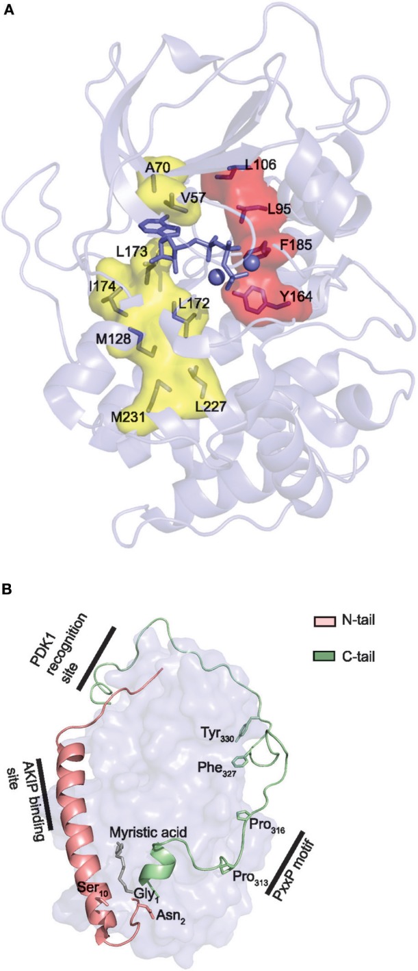
Core and tail structures of the PKA C subunit. (A) C- and R-spines in Cα1. In the active conformation of kinases, the C- and R-spines are assembled. In the case of PKA Cα1, the residues constituting the C-spine (yellow) are Ala70, Val57, Leu173, Ile174, Leu172, Met128, Met231, and Leu227. The adenine nucleobase of ATP (slate stick presentation) is also part of the C-spine. The R-spine (red) consists of Cα1 residues Leu106, Leu95, Phe185, and Tyr164. One-letter aa abbreviations are used in the figure. Figure is based upon (175). PDB identifier 3FJQ (169). (B) Presentation of the conserved kinase core (rendered as a surface in slate) of Cα1, including the N-tail (salmon) and C-tail (green) in cartoon presentations. Myristic acid (gray) is shown bound to the hydrophobic pocket. Selected structures and residues in the N- and C-tails are highlighted and described in the text. PDB identifier 1CMK (73).