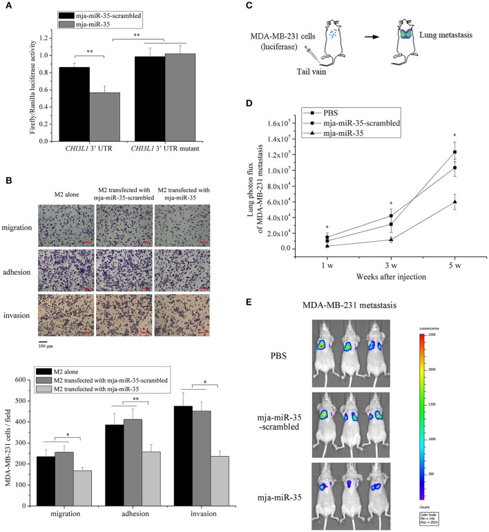
Figure 3.
Underlying mechanism of mja-miR-35-mediated suppression of breast cancer cell metastasis. (A) The direct interaction between mja-miR-35 and CHI3L1 3′ UTR. The 293T cells were co-transfected with mja-miR-35 and pmir-GLO vector fused with CHI3L1 3′ UTR. Firefly and Renilla luciferase activities were determined. As controls, mja-miR-35-scrambled and CHI3L1 3′ UTR mutant were included in the co-transfections (**p < 0.01). (B) The impact of mja-miR-35 expression in M2 macrophages on breast cancer cell metastasis. MDA-MB-231 cells were co-cultured with mja-miR-35-transfected M2 macrophages, followed by cell migration, adhesion and invasion assays (*p < 0.05; **p < 0.01). Scale bar, 100 μm. (C) Schematic diagram of MDA-MB-231 cell metastasis to lung following intravenous injection in mouse. (D) The effects of mja-miR-35 on breast cancer cell metastasis in vivo. MDA-MB-231 cells were intravenously injected into BALB/c nude mice. One day later, mja-miR-35, mja-miR-35-scrambled or PBS was injected into the mice. Lung metastasis was monitored every 2 weeks (*p < 0.05). (E) Representative images of MDA-MB-231 cell metastasis in mice at week 5.

