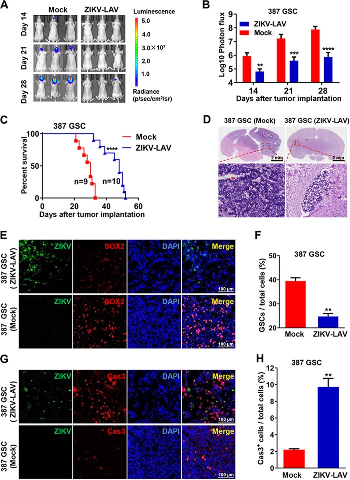
FIG 2.
ZIKV-LAV inhibited GSC tumor growth and prolonged animal survival. (A to C) Three-week-old BALB/c nude mice were intracerebrally transplanted with 50,000 luciferase-labeled type 387 GSCs mixed with 10,000 PFU of ZIKV-LAV or RPMI 1640 (Mock). (A) In vivo bioluminescence imaging of tumor growth from luciferase-labeled 387 GSCs at the indicated times. (B) Quantification of photon flux from ROI analysis of the dorsal side. Statistical significance was analyzed by unpaired Student’s t tests. **, P < 0.01; ***, P < 0.001; ****, P < 0.0001. (C) Survival curves. Statistical significance was determined by the log rank test. ****, P < 0.0001. (D to H) Three-week-old BALB/c nude mice transplanted with 387 GSCs mixed with ZIKV-LAV or RPMI 1640 (Mock) were sacrificed on day 23 postimplantation. The brain tissues were collected and prepared for cryosectioning. (D) Representative images of brain sections stained with hematoxylin and eosin. The tumor areas are enclosed with black lines. (E) Immunofluorescence staining of cryosections for the ZIKV E protein (green), SOX2 (red), and DAPI (blue). The percentages of GSCs in tumor tissues are shown as means ± SD (n = 3; right panel). **, P < 0.01. (F) Cleaved caspase 3 (Cas3) staining of apoptotic cells in tumor tissues. The percentages of Cas3+ cells in tumor tissues are shown as means ± SD (n = 3; right panel). **, P < 0.01.

