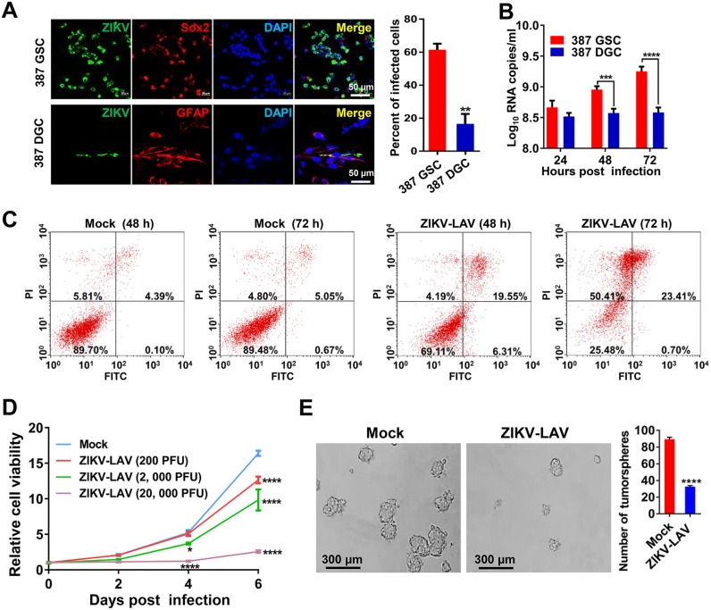
FIG 4.
ZIKV-LAV preferentially infects and kills GSCs and impairs tumorsphere formation. (A) Immunofluorescence staining of ZIKV-LAV-infected 387 GSCs and DGCs for viral E protein (green), SOX2 or GFAP (red), and DAPI (blue) on day 3 postinfection (MOI = 0.1). (Right panel) Percentages of infected cells. Statistical analysis was performed using unpaired Student’s t tests. **, P < 0.01. (B) Growth curves of ZIKV-LAV in 4121 GSCs and DGCs (MOI of 0.1). Viral RNA copies in culture medium were analyzed by RT-qPCR at the indicated time points. Statistical analysis was performed by two-way ANOVA. ***, P < 0.001. ****, P < 0.0001. (C) Analysis of cell death induced by ZIKV-LAV infection. Briefly, 387 GSCs were infected with ZIKV-LAV (MOI of 1), collected at the indicated time points, and subjected to the cell death assay by flow cytometry. (D and E) GSCs were seeded in 96-well plates at a density of 2,000 per well, immediately infected with the indicated dose of ZIKV-LAV, and subjected to cell viability analysis using the Cell Titer-Glo assay at the indicated time points (D) and the tumorsphere assay on day 5 postinfection (E). Statistical analysis was performed by two-way ANOVA. *, P < 0.05; ****, P < 0.0001.

