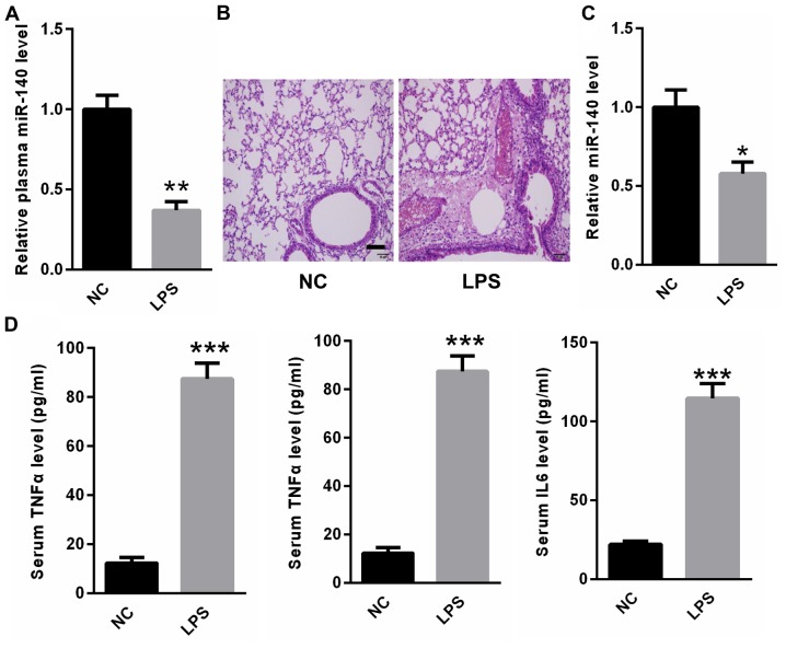
Figure 3.
miR-140 expression was lower in the peripheral blood and lung tissues of rats with LPS-induced ALI. (A) miR-140 was decreased in the peripheral blood of rats in the LPS group compared with NC rats. (B) Hematoxylin and eosin staining revealed that rats in the LPS group developed obvious lung inflammation, hemorrhaging and alveolar septal thickening compared with the NC group. Scale bar=25 µm (C) miR-140 expression was decreased in the lung tissues of LPS rats compared with NC rats. (D) ELISA results revealed that serum levels of TNF-α, IL-6 and IL-1β were significantly increased in ALI rats compared with NC rats. *P<0.05, **P<0.01 and ***P<0.001 vs. NC. miR, microRNA; LPS, lipopolysaccharide; ALI, acute lung injury; NC, negative control; TNF, tumor necrosis factor; IL, interleukin.

