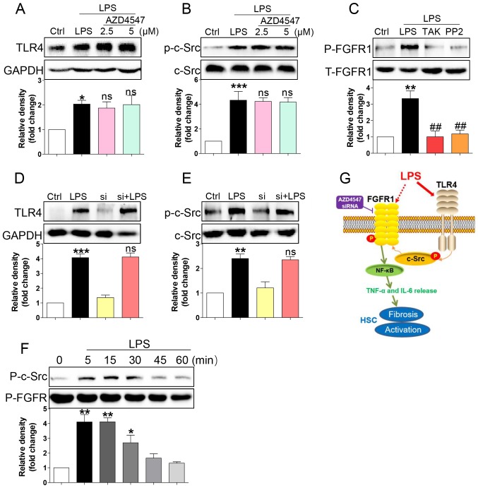
Figure 4.
LPS triggers FGFR1 phosphorylation in HSCs through the TLR4/c-Src signalling cascade. HSCs were pre-treated with AZD4547 (AZD, 2.5, 5 µM) for 1 h. After LPS treatment for 15 min, the expression of (A) TLR4 and (B) phosphorylation levels of c-Src were detected by western blotting (WB). (C) HSCs were pre-treated with TAK-242 (TLR4 inhibitor), or PP2 (c-Src inhibitor) for 1 h, followed by LPS treatment for 15 min. Phosphorylation of FGFR1 was determined via WB. (D and E) siFGFR1 did not reduce LPS (15 min)-induced increase expression of TLR4 and c-Src activation. (F) LPS-induced interaction between c-Src and FGFR1. HSCs were exposed to LPS for the indicated times. Lysates were then subjected to c-Src IP and FGFR1 was measured. (G) Schematic illustration of the major findings of this study: LPS activated TLR4 and increased signalling through the c-Src pathway to cause FGFR1 activation and production of inflammatory cytokines. This proinflammatory response produced HSCs activation and liver fibrosis (*P<0.05, **P<0.01, ***P<0.001, vs. Ctrl group; ns, not significant vs. LPS group; ##P<0.01, vs. LPS group).

