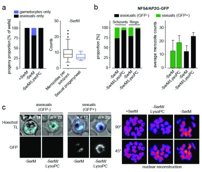
Figure 3. Progeny analysis in single cells.
( a) Analysis of progeny from 96 single cells in the Pf2004/164tdTom reporter line (expressing tdTomato reporter under the gametocyte-specific PF10_0164 promoter 17). Each parasite produces only asexual or gametocyte progeny (left panel). The number of sexual progeny corresponds to the number of daughter merozoites per schizont under lysophosphatidylcholine-deficient medium (−SerM) conditions (right panel). A total of 50 independent measurements were made per condition. ( b) Analysis of sexually committed schizonts in the NF54/AP2-G GFP reporter line. The proportion of GFP positive schizonts and the number of GFP positive progeny (rings) correlate and mark sexual cells (gametocyte rings). Both sexually committed (GFP-positive) and asexually committed schizonts show reduced merozoite number under −SerM conditions (right bar graph). GFP-positivity was quantified in 100`000 cells by ImageStream analysis in biological triplicates. Merozoite counts represent the mean of 100 measurements per condition, performed in biological triplicates. ( c) Representative images from cells in ( b) and selected planes from 3D reconstructions of nuclei are shown in the left and right panels, respectively. Hoechst, nucleic acid stain; TL, transmitted light. n = number of nuclei.

