
Figure 4.
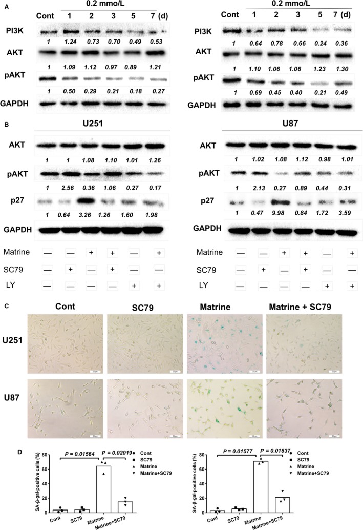
Matrine inhibits the PI3K/AKT/p27 signaling pathway. A, Western blot analysis for PI3K, AKT, pAKT, and GAPDH protein levels in protein lysates (20 μg) prepared from U251 and U87 cells treated with matrine for the number of days indicated. B, Western blot analysis to detect levels of AKT, pAKT, P27, and GAPDH in lysates prepared from U251 and U87 cells treated with matrine (0.2 mmol/L), AKT activator SC79 (5 μg/mL), or PI3K inhibitor LY294002 (1 mmol/L) 72 h. C, In situ SA‐β‐gal assay to detect senescent cells. Cells were treated with 0.2 mmol/L matrine and AKT activator SC79 for 72 h. Scale bars = 20 μm. D, Graphic representation of the percentage of SA‐β‐gal‐positive cells determined in four random fields per sample. All data are expressed as the mean ± SD of values from experiments performed in triplicate. Statistical analyses were performed using permutation test. P‐value came from the comparison between treated group and control group or between two treated groups