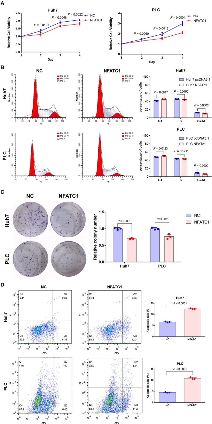
Figure 3.

NFATc1 inhibits HCC cell proliferation and induces apoptosis. Huh7 and PLC HCC cell lines were transfected with NFATc1 overexpression plasmid (NFATc1) or vector plasmid for negative control (NC). A, Ectopic expression of NFATc1 inhibited proliferation of Huh7 and PLC cells, as shown by CCK‐8 assay. The experiments were performed in quadruplicate wells three times. Data are presented as the mean ± SD. B, Ectopic expression of NFATc1 caused G1 arrest in Huh7 and PLC cells. Images of flow cytometry analysis are shown on the left, and the mean ± SD of cell percentage for each group is shown on the right. The experiments were performed in triplicate wells three times. Data are presented as the mean ± SD. Dots represent data from cells in triplicate wells under the same treatment. C, NFATc1 inhibited colony formation in Huh7 and PLC cells. Images are shown on the left, and the mean ± SD of relative colony number for each group is shown on the right. The experiments were performed in triplicate wells three times. Data are presented as the mean ± SD. Dots represent data from cells in triplicate wells under the same treatment. D, NFATc1 induced apoptosis in Huh7 and PLC cells as shown by flow cytometry following Annexin V‐APC and PI staining. The experiments were performed in triplicate wells three times. Data are presented as the mean ± SD. Dots represent data from cells in triplicate wells under the same treatment. Statistical analysis for all the experiment data in Figure 3 was performed using two‐tailed Student t tests
