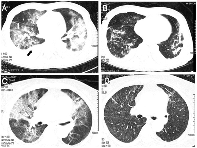
Figure 1.
Results of chest computed tomography scans. (A) Chest computed tomography revealed GGO (white arrow) of lung fields with pleural effusion (black arrow) in Case 1. (B) GGO dissapearedfollowingtreatment of case 1 with caspofungin. (C) GGO were found in both lungs in Case 2. (D) GGO of case 2 were absorbed followingtreatment with caspofungin. GGO, ground-glass opacity.

